* This product is for research use only. Not intended for use in the treatment or diagnosis of disease.
Electroporation is the most effective non-viral gene delivery method for introducing DNA, RNA, mRNA, RNP, proteins, and other molecules into a variety of cells (especially cells that are difficult to transfect, such as primary and stem cells). By using a precise pulse current, it can induce transient pores in the phospholipid bilayer of the cell membrane, so that the cell can absorb extracellular genetic material.
Studies have shown that during electroporation, the increased transmembrane voltage plays two roles: (a) forming pores and (b) providing local driving force.
During the opening of the pore, the nucleic acid can enter the cell and eventually enter the nucleus. Linear DNA with free ends has higher recombination capacity and is more likely to integrate into the host chromosome to produce stable transformants. Supercoiled DNA is more easily packaged into chromatin and is usually more effective for transient gene expression.
Electroporation is used in the fields of molecular biology research and medicine.
Electroporation can be used to transfer a variety of genetic material into cells, including DNA, RNA, and oligonucleotides (including synthetic oligonucleotides with uncharged backbones), and to transfer plasmids directly between cells.
In vivo electroporation has been shown to be effective for a variety of tissues, including tumors, skin, liver, lung, kidney, thymus, bladder, adipose tissue, vasculature, retina, cornea, ciliary muscle, brain, the umbilical cord of the spinal cord, skeletal muscle, and testis. It can be used to deliver a series of genetic material, such as DNA, RNA, and oligonucleotides (e.g., siRNA, antisense oligonucleotides).
Now, in vivo electroporation has been used for transdermal drug delivery, cancer tumor electricity chemotherapy, and the effective delivery of gene therapy and vaccines.
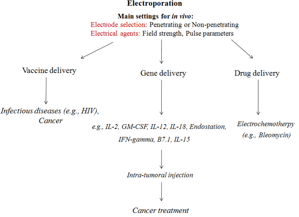 Figure 1: Common applications of electroporation
Table 1: Brief of electroporation
| What can be electroporated? | Typical transfectants |
|---|---|
| Bacteria Fungus/yeast Plants Others, insects, fish, mold and amphibians Mammalian Primary explant culture Established cell line Human, in vitro, in vivo and ex vivo | DNA/RNA Antibodies/proteins Drugs Other molecules/ions |
Optimize electroporation conditions for each cell type to ensure successful results. The following suggestions can help produce high-efficiency electroporation for most cell types.
According to the purpose of the experiment, the incubation time is determined by the nature of the plasmid and the half-life of the expressed protein.
Reference